
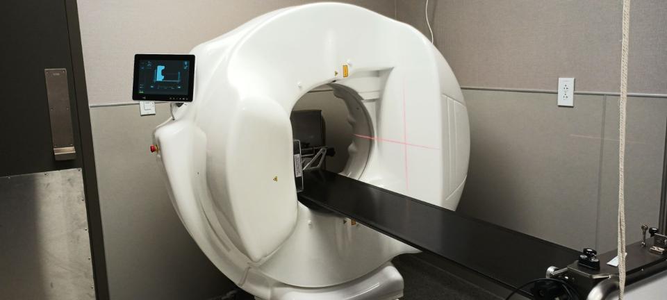
What Is Vimago?
The Vimago system is similar to a Computed Tomography (CT) scanner, but has a patented High Definition Volumetric Imaging (HDVI) technology that creates high resolution, detailed images in both 2D and 3D with very little radiation.
This technology allows for greater detail and allows for improved diagnostic imaging, making it easier and faster to diagnose, and thus treat patients. The Vimago images of the skull (teeth and sinuses), bones (joints, backs), chest (lungs), abdomen (vasculature), and soft tissue or bony masses can be viewed in both 3D with the layers of the body removed electronically for easier visualization, or in the traditional 2D circular view.
The unit also has built-in Fluoroscopy technology which creates a real-time radiograph in motion and can be used for many applications. For more information, call the office, or stop by for a tour!
Reasons Vimago Imaging is Recommended:
- Dentistry
- Skull / Sinuses
- Tumors / Masses – identification and assessment
- Trauma
- Metastasis to chest and abdomen
- Spinal / Joint abnormalities
- Surgical planning
This technology is only available at the Care First Animal Hospital at Oberlin location.
Vimago Anatomy Guide
Anatomy included in Vimago HDVI Scan
- Skull—Tip of nose through caudal fossa. Both pre and post-contrast:
- Dental
- Ear bulla
- Nasal/sinuses
- Orbit
- TMJ
- Brain with Contrast
- Cervical Spine—Caudal Fossa thru T-3
- Bone/soft tissue
- Esophagus/trachea
- Thyroid
- Thoracic spine—Body of T1 thru T13
- Lumbar Spine—Body of T13 thru S3
- Thoracic region—Chest wall, Heart, Lungs, and Mediastinum. Patients will have a non-contrast study and if indicated a post-contrast study.
- Abdominal region—Abdomen, Adrenals, GI tract, Kidneys, Liver, Pancreas, Spleen, and Vascular system. Patients will have a non-contrast study and if indicated a post-contrast study.
- Portosystemic shunt with contrast
- Ectopic Ureter with contrast
- Pelvic region—Bladder, Pelvis, and Prostate. Exam individualized to the patient based on diagnostic imaging studies, history, and symptoms
- Joints—The joint and connecting soft tissue structures.
- Carpi, Elbows, Shoulder, Tarsi, Stifle, Hips, Paws, Long bones
- Soft tissue mass, tumor—Individualized to each patient to include all boarders of the mass. Both pre and post-contrast studies.
Anatomy included in Vimago Fluoroscopy
- Cough assessment
- Esophagram/Barium Swallow
- Gastrogram
- Upper GI
- Barium Enema
- Excretory urogram